


Neuroimaging and Neurointervention
Neuroimaging
Neuroimaging has become an indispensable part of non-invasive assessment of brain and spine disorders. Depending on patients’ symptoms and clinical suspicion, various types of neuroimaging modalities will be employed, including computer tomography (CT), magnetic resonance imaging (MRI), single proton emission computer tomography (SPECT), positron emission tomography (PET), and catheter angiography.
Magnetic Resonance Imaging
MR Brain
MR Spine
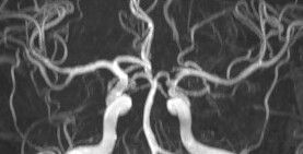
MR Angiogram
- Assess presence and degree of narrowing of intracranial arteries
- Useful in evaluating intracranial aneurysms and arteriovenous malformation
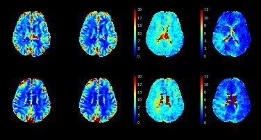
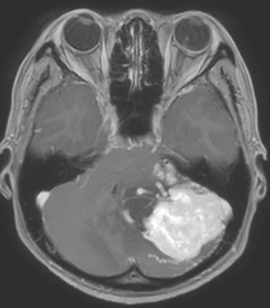
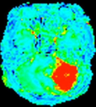
MR Perfusion
By imaging contrast passage through brain parenchyma, the status of microcirculation of the brain can be assessed
- MR perfusion has been employed to assess malignant potential of brain tumour, as well as to follow up treatment response
- MR perfusion study performed with Diamox challenge is useful in assessing degree of cerebrovascular reserve in chronic steno-occlusive disease
- MR perfusion is useful in assessing the amount of salvageable brain tissue in acute ischemic stroke
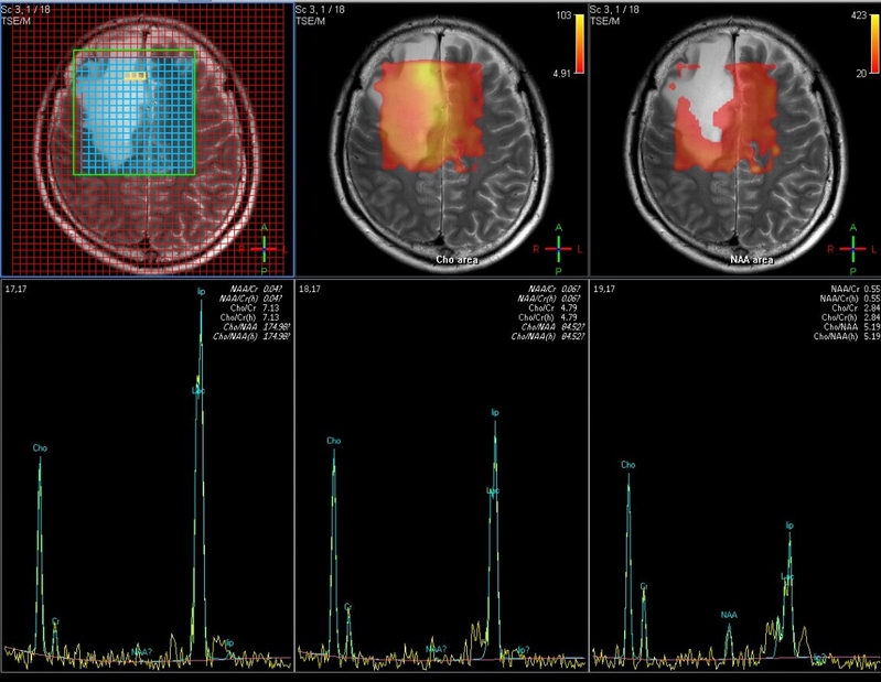
MR Spectroscopy
- Different metabolites of the brain tissue show slightly different MR signal, which can be picked up by MR spectroscopy
- MR spectroscopy has been employed to evaluate the malignant potential of brain tumour, as well as to follow up treatment response
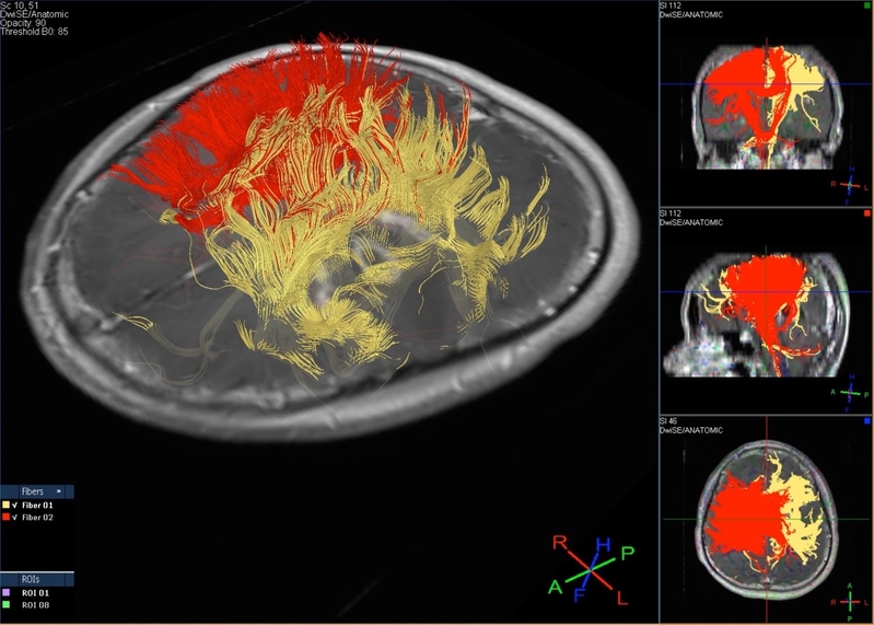
MR Diffusion Tensor Imaging and Tractography
- Densely packed fiber tracts of the brain show restricted diffusion of its water molecule, and hence can be imaged using diffusion tensor imaging
- Diffusion tensor imaging and tractography can map major fiber tracts of the brain for surgical planning, in order to allow safe brain tumour or epilepsy surgery
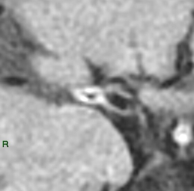
Vessel Wall Imaging
- High resolution MRI is now capable of imaging the thin vessel wall, which is otherwise not visualised in conventional imaging modalities
- It allows assessment of vessel wall status to determine the etiology of vessel narrowing, such as atherosclerosis, vasculitis, etc.
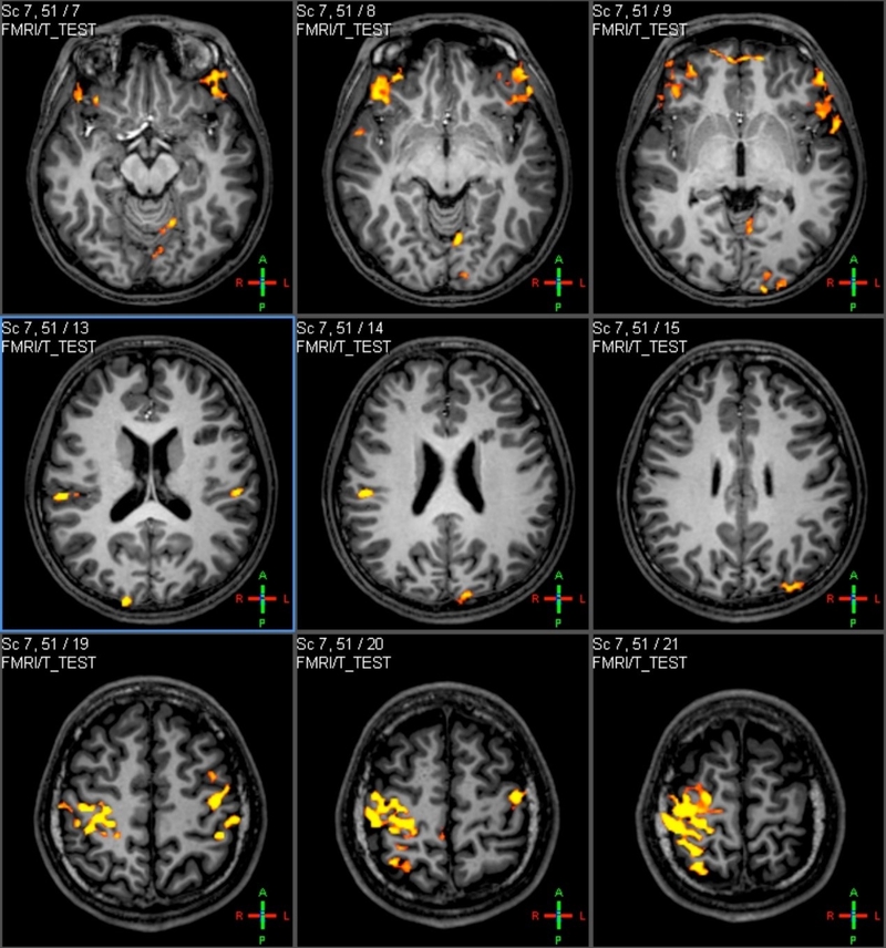
Functional MRI
- During motor or language task, there will be increase in blood flow to the functional cortex of the brain. Such change in blood flow can lead to subtle change in MR signal, which can be picked up and used to localise the motor or language area
- Functional MRI is an important pre-surgical planning tool for brain tumour or epilepsy surgery
Neurointerventional Procedure
With the advancement of angiographic technology and medical devices, more and more neurovascular diseases can now be treated minimally invasively by endovascular approach. These include cerebral aneurysms, arteriovenous malformation / fistula, arterial stenosis, etc. Neurointerventional procedures are also useful in preoperative embolisation of head and neck tumour or in stopping bleeding in the head and neck region. Neurointerventional procedures can be broadly divided into those aiming at imaging of vessels (diagnostic angiogram), occlusion of vessels (embolisation) or opening up of narrowed / occluded vessels (revascularisation).
Diagnostic angiogram
Most neurointerventional procedures start with a diagnostic angiogram. This is done by puncturing one of the arteries in the groin (femoral) or wrist (radial) by a fine needle. By threading a small wire through the needle into the artery, a plastic tube (sheath) can be inserted. Various catheters and guidewires can then be advanced into the head and neck arteries, from which contrast can be injected under X-ray guidance to obtain diagnostic images.
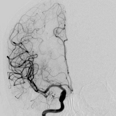
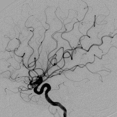
Catheter cerebral angiogram
Catheter spinal angiogram
Intravenous cone beam CT angiogram
Embolisation
Various conditions, such as aneurysms, arteriovenous malformation / fistula, tumour, bleeding, etc, can be treated endovascularly by occluding the supplying arteries. This can be done by introducing a very small tube (microcatheter) into the supplying artery, where different kinds of materials (embolic agents) can be injected / deployed to occlude the vessels. Depending on the pathology, the embolic agents used include coils, particles, tissue glue, liquid polymer, etc.
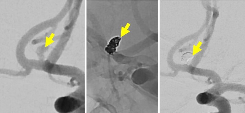
Embolisation of intracranial aneurysm
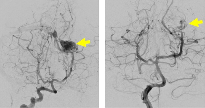
Embolisation of intracranial arteriovenous malformation / fistula
Embolisation of intracranial dissection
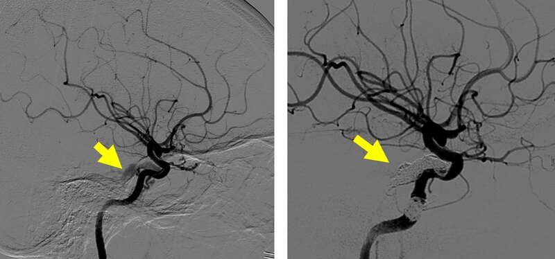
Embolisation of carotid-cavernous fistula
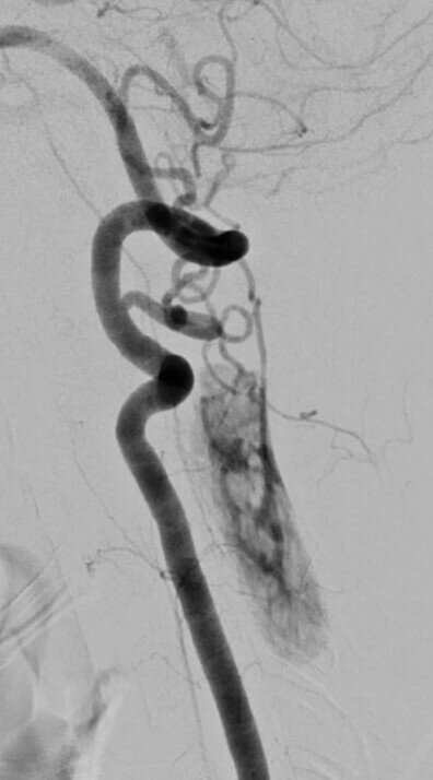
Embolisation of intracranial / head and neck tumour
Embolisation for head and neck bleeding
Embolisation of spinal arteriovenous malformation / fistula
Embolisation of spinal tumour
Revascularisation
Narrowing of the head and neck arteries is an important cause of ischemic stroke, which can be treated by open surgery or endovascular approach. A narrowed artery can be opened up endovascularly using a balloon (angioplasty) or metallic stent (stenting). With the introduction of embolic protection device, the safety of angioplasty and stenting of the head and neck vessels have been markedly improved.
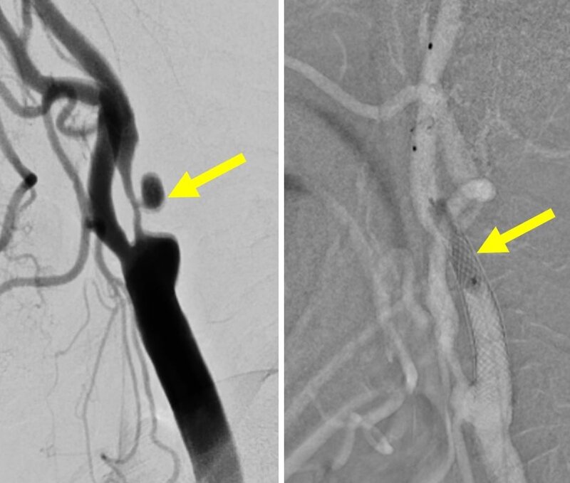
Angioplasty / stenting of extracranial carotid artery
Angioplasty / stenting of extracranial vertebral artery
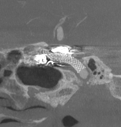
Angioplasty / stenting of intracranial arteries
Angioplasty / stenting of intracranial veins
Miscellaneous
Balloon occlusion test
Wada’s test
Intra-arterial infusion of chemotherapy
Inferior petrosal sinus sampling